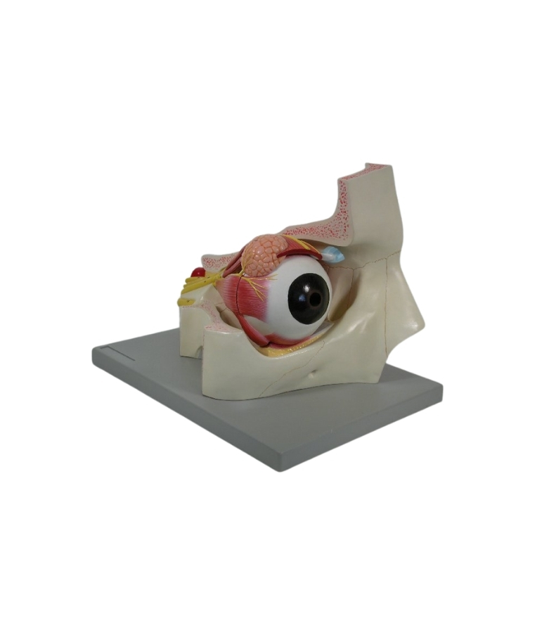
This anatomical model depicts the human eye with the optic nerve situated in its natural position within the bony orbit, including the floor and medial wall. It can be dissected into the following parts:
Both halves of the sclera with optic nerve and eye muscles
Cornea
Lens
Vitreous humour
Superior rectus muscle ( rectus superior)
Lateral rectus muscle ( rectus lateralis)
Mounted on a base. Dimensions: 19 ? 20 ? 28 cm Weight: 1.5 kg.


Reviews
There are no reviews yet.