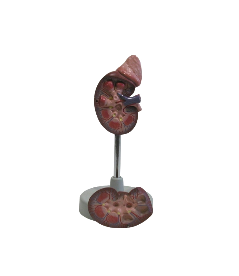
This life-size kidney model illustrates the development and typical locations of kidney stones (nephrolithiasis) and urinary stones (urolithiasis). The right kidney is sectioned open to show the renal calices, pelvis, and ureter, with stones positioned in the:
In the area of the renal pyramids
In the area of origin of the upper calix group
In the renal cortex
In the connecting tubule of the lower calix group, causing congestion of the minor calices (partially closed, partially opened)

Reviews
There are no reviews yet.