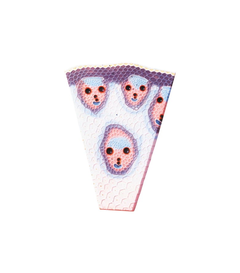
This model provides a clear, three-dimensional view of the anatomical organization and tissue differentiation in a monocot stem, aiding students in understanding its unique characteristics.The model vividly illustrates the epidermis, hypodermis, ground tissue, vascular bundles, xylem, phloem, and sclerenchyma. It highlights the scattered arrangement of vascular bundles, which is a key identifying feature of monocot stems. Each vascular bundle is shown with xylem vessels, phloem tissues, and bundle sheath cells, along with parenchymatous ground tissue that fills the entire stem section. Made from high-quality, durable fiber material, the model is hand-painted in distinct colors to differentiate each tissue type and structure for visual clarity. It is mounted on a sturdy base and includes a numbered English key card to help in the identification and explanation of each component during classroom instruction.

Reviews
There are no reviews yet.