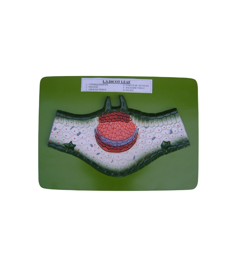
Representation showing the longitudinal section of a dicot leaf, designed for use in biology and botany laboratories. This educational model effectively demonstrates the internal anatomy and tissue organization of a dicot leaf, making it an essential tool for teaching plant structure and function. The model clearly illustrates the key components of a dicot leaf, including the upper and lower epidermis, palisade and spongy parenchyma tissues, vascular bundles with xylem and phloem, and stomata with guard cells. The vascular bundles are prominently shown within the mesophyll region, helping students visualize the pathway of water, nutrients, and photosynthates. Constructed from high-quality, unbreakable fiber material, the model is hand-painted in contrasting colors to distinctly highlight various tissues and cellular structures. It is mounted on a durable base and includes a numbered English key card for easy identification and explanation of each part.

Reviews
There are no reviews yet.