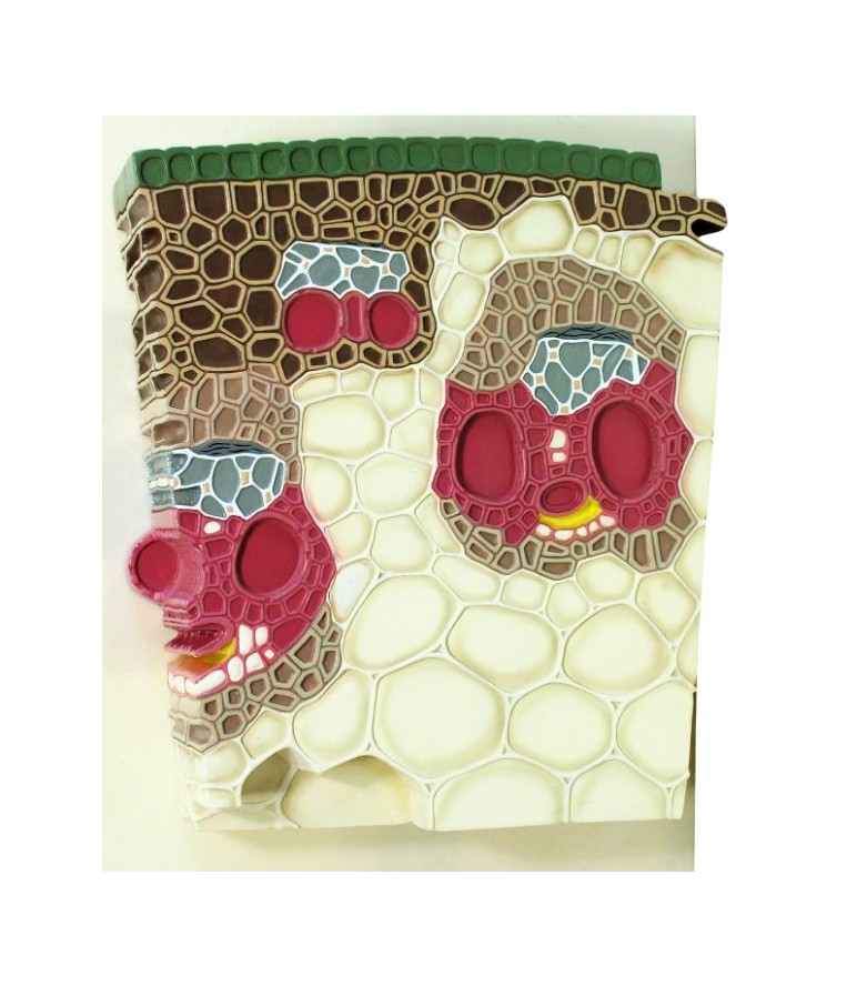
The model provides a clear and comprehensive view of the cellular organization and tissue differentiation found in the stem or shoot of woody plants. This model accurately depicts the epidermis, cortex, vascular bundles (xylem and phloem), cambium, and pith regions, showing their arrangement and interrelationship. The xylem vessels, tracheids, and sieve tubes are highlighted in distinct colors to represent the pathways for water and nutrient transport. The cambial zone is clearly illustrated to emphasize its role in secondary growth, while the epidermal layer demonstrates the presence of protective tissues. Constructed from high-quality, unbreakable fiber material, the model is hand-painted in vibrant, contrasting colors to differentiate each tissue type for easy recognition. It is mounted on a durable base, making it ideal for classroom display and long-term use. The model comes numbered with an English key card for identification and reference of all major components.
Reviews
There are no reviews yet.