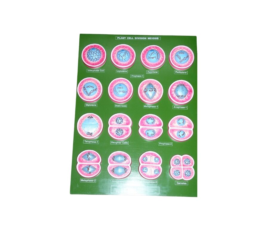
This set includes 12 beautifully coloured models illustrating the detailed microscopic structure observed during the stages of meiotic cell division. Each model vividly represents a specific stage, including leptotene, zygotene, pachytene, diplotene, diakinesis, metaphase I, anaphase I, telophase I, prophase II, metaphase II, anaphase II, and gamete formation. All models are securely mounted on a base, with parts clearly numbered and accompanied by an English key card for easy identification and study.
Reviews
There are no reviews yet.